Advanced Bioimaging
The Advanced Bioimaging Unit (UBA) is a joint unit between the University of the Republic and Institut Pasteur de Montevideo. We are passionate about the development of new microscopy hardware and methods, and have a strong commitment to the dissemination of our tools. At UBA we offer the region free microscopy workshops on advanced techniques and methods, as well as practical analysis courses and microscopy fundamentals for beginners.
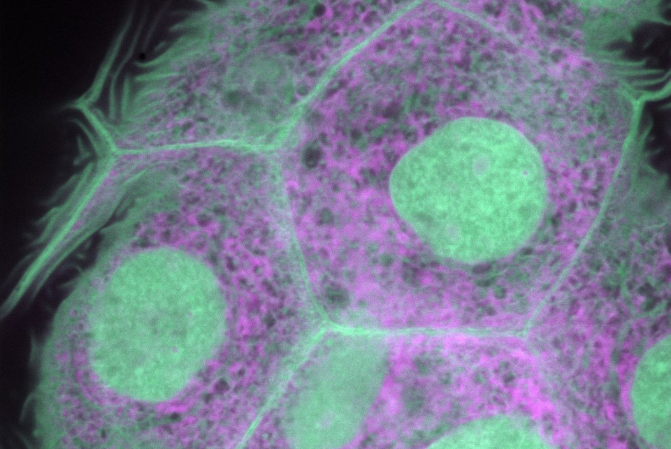
UBA’s staff of expert microscopists have solid experience in supporting users in the design, execution, and troubleshooting of microscopy experiments, and we provide services following international standards of good practice. UBA’s microscopy portfolio includes epifluorescence, confocal laser instruments, as well as “custom-made” multiphoton instruments. Our microscopes can perform experiments up to 5D (x / y / z / t / λ o τ). Among the tools that we master at the UBA, techniques such as FLIM, FRET, phasor graphs, autofluorescence, harmonic generation, deep tissue imaging, FCS and related techniques stand out, among others.
Members
Bruno Pannunzio, PhD

Research lines
We seek to provide public access to high-quality bioimaging services, and to develop instrumentation, methods and applications in biophotonics and bioimaging focused on life sciences and more specifically focused on biomedical research.
Technology Driven Research (TDR): innovative Instruments and Methods.
Spectroscopy imaging tools and methodsfor in vivo metabolic imaging using label-free approaches.
Development and application of methods based on phasor graphs for hyperspectral and lifetime fluorescence imaging.
Biology Driven Project (BDP): Strongly supported by collaborations (intra and inter-institutional).
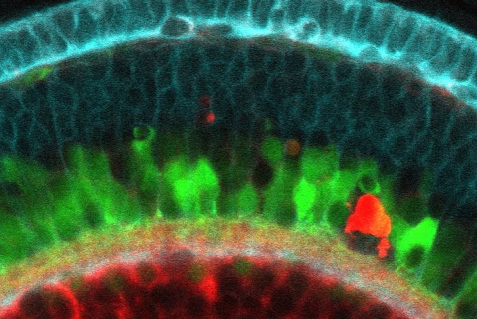
In vivo water dynamics using DAN probes and images. Focus on neurodegenerative diseases and liquid phase separation.
Metabolic signatures based on autofluorescence molecules for imaging pathology.
Main equipment
COMMERCIAL EQUIPMENT IN SERVICE
Epifluorescent microscopy:
Olympus IX81 Epifluorescence Microscope: inverted microscope with Halogen and Mercury illumination that has a Hamamatsu ORCA ER monochrome camera for the acquisition of very good quality fluorescence and transmitted light images.
Applications:
- Observations of slides, petri dishes, bottles and other types of samples labeled with fluorescent molecules.
- Observations of preparations in bright field, phase contrast and DIC (Nomarski).
- Taking images with an ORCA ER Hamamatsu monochrome camera.
Confocal microscopy:
Zeiss LSM800 confocal: Axio Observer Z1 inverted epifluorescence microscope coupled to LSM800 confocal module. It is a basic semi-spectral confocal that allows the acquisition of high-resolution fluorescent confocal images in a wide variety of samples. It also has an AxioCam 506 monochrome camera to obtain fluorescence, bright field or DIC images in parallel.
Applications:
- Acquisition of confocal images of fluorescent samples in a plane or in depth.
- Imaging with transmitted light and DIC that can be combined with fluorescence.
- Acquisition of fluorescent, brightfield or DIC images with a high-speed monochrome camera.
- Carrying out experiments that involve taking images from various positions in 2D or 3D, mosaic-type reconstructions, time-lapse, semi-spectral scanning, among others.
Zeiss LSM880 confocal: Axio Observer 7 inverted epifluorescence microscope coupled to LSM880 confocal module. It is a spectral confocal that has a high sensitivity GaAsP detector. Fluorescent or transmitted light images can be obtained with DIC of excellent definition. It is an instrument equipped with an incubation chamber with temperature and C02 controlled for in vivo experiments.
Applications:
- Acquisition of confocal images of fluorescent samples in a plane or in a volume.
- Imaging with transmitted light and DIC that can be combined with fluorescence.
- Carrying out experiments that involve taking images from various positions in 2D or 3D, mosaic-type reconstructions, time-lapse, semi-spectral scanning, among others.
- Realization of experiments with living samples for long periods of time with focus stabilization.
- Spectral acquisitions.
- Fluorescence correlation spectroscopy (FCS).
COMPUTERS FOR IMAGE PROCESSING
Computer room UBA-IPMONT:
UBA02 (IPMONT):
- 22” and 28” screens.
- i5 processor.
- RAM 16 GB.
- Analysis programs: , Fiji & ImageJ (NIH, Bethesda-USA), SimFCS for images (G-Soft, Ilinois-USA)/Zen Lite blue-black (Carl Zeiss Microscopy, Munich-Germany).
UBA03 (IPMONT):
- 28” screen and interactive drawing monitor, XP-PEN Artist 22E Pro 21.5”.
- i7 processor.
- RAM 64 GB.
- Analysis programs: , Fiji & ImageJ (NIH, Bethesda-USA), SimFCS for images (G-Soft, Ilinois-USA)/Zen Lite blue-black (Carl Zeiss Microscopy, Munich-Germany).
Computer room UBA-HC:
PC-1 (HC):
- Intel Core i7-3779 CPU @ 3.40HGHz.
- 32Gb DDR3 RAM.
- 128GB SSD, HDD SATA 1Tb.
- NVIDIA GeForce GT 640 DDR3 2Gb video card.
- 2 BenQ GL2450 26-inch monitors.
PC-2 (HC):
- CPU INTEL CORE I7 9700 (3.0Ghz) 12MB Socket 1151.
- 64Gb DDR3 RAM.
- HD SSD 240GB KINGSTON, HD. 1TB SATA6.
- 2 monitors LED MONITOR 27″ ACER Nitro VG270 IPS 144Hz FULL HD.
SYNOLOGY DS920+ DESKTOP NAS data server (4 BAYS) with 40TB of data.
Software for image analysis and processing:
- SimFCS for images (G-Soft, Ilinois-USA), Fiji & ImageJ (NIH, Bethesda-USA), Icy (Pasteur Paris, France) y código/rutinas customizadas en MatLab (MathWorks).
Workshop equipment and measuring devices (IPMONT):
- 3D printer – Comgrow Creality Ender 3 Pro.
- Grizzly G0602 Metal Lathe – 10″ x 22″.
- HiTorque Mini Mill, Deluxe Metal Milling Machine (LittleMachineShop.com).
- Accessories for milling and lathe (angle clamp and rotary).
- YIHUA 862BD+SMD ESD multi-function hot air soldering station °F /°C.
- Compact Power and Energy Meter Console, Digital 4″ LCD + Microscope Slide Thermal Power Sensor, 300 nm – 10.6 μm, 2 W.
- Tektronix TDS784A digital oscilloscope (4 channels 1GHz).
- High resolution and speed spectrometers for diffuse optical spectroscopic imaging (DOSI). HR4000 y 2x USB200 (Ocean Optics).
EQUIPMENT BEING DEVELOPED
Fiber-optic based instrumentfor lifetime and spectral measurements. Hamamatsu 2×2 detector, A320 fastFLIMbox (ISS), Diode Laser 370 nm modulated up to 300MHz (ISS), Fiber optics (ThorLabs).
Two-photon home-built microscope with lifetime resolution and FCS. Nikon Ti-S epifluorescent block, fiber-based pulsed laser 780 nm (Calmar), 3 pseudo-photon counting detectors (GaAsP Hamamatsu), FLIMBox 320 (ISS), motorized x/y stage (ASI), electric lens (Optotune), CO2 and temperature controlled, optical table with passive isolation (TMC) x/y galvanometric scanner (Cambridge Technologies).
- FLIM
- FRET
- Phasor plots
- FCS (single point, line, circular, diffusion map)
- RICS (crossRICS)
- N&B (crossN&B)
- pCF (pCOMB)
- 2D y 2D Particle Tracking
- Epifluorescence (camera based)
2-photon microscope with DIVER detector (1 or 4 channels) + 2 non-scanning detectors for epifluorescence (deep tissue/intravital imaging). Tunable Pulsed Laser (690-1040) Ti-Sapphire MaiTai eHP DeepSee (Spectra-Physics), 2-inch scanning optics, 2 pseudo photon counting detectors + custom DIVER detector (GaAsP Hamamatsu), FLIMBox Kintex (FLIM Labs), stage motorized x/y/z (ASI), electric lens (Optotune), optical table with passive isolation (NewPort), x/y galvanometric scanner (Cambridge Technologies).
- FLIM
- FRET
- Phasor plots
- FCS (single point, line, circular, diffusion map)
- RICS (crossRICS)
- N&B (crossN&B)
- pCF (pCOMB)
- 2D y 2D Particle Tracking
- Spectral detection (sine/cosine filters)
Courses
- 1º Annual Workshop on Advanced Microscopy and Biophotonics. November 25 – 28, 2019. Advanced and Biphotonic Microscopy Unit, Hospital de Clínicas – University of the Republic. Organizer: Malacrida, Leonel.
- 2º Annual Workshop on Advanced Microscopy and Biophotonics. November 23 – 27, 2020. Institut Pasteur de Montevideo / University of the Republic. Organizer: Malacrida, Leonel.
User training:
- Basic use of fluorescent microscopy instruments (one to one). Díaz, Marcela.
- Management, storage and analysis of basic data using free software (one by one). De los Campos, Tabaré.
Projects
1- Projects driven by the development of technology, methods and applications in the field of Biophotonics/Technological Driven projects.
a) «Development and fine-tuning of a new device based on optical fibers with lifetime and spectral resolution for in vivo tissue metabolic evaluation by autofluorescence.»funded by CSIC I+D 2021-2023. PI: Leonel Malacrida. ~40K (U$S)
b) “Two-photon microscopy resolved in lifetime at the University Hospital: a state-of-the-art microscope to answer questions in biomedicine.”, funded by CSIC Heavy Equipment 2018. PI’s: Leonel Malacrida and Javier Hurtado. $U 3.850.000
c) “Development of an optical device for the rapid and portable diagnosis of SARS-CoV2 with engineered proteins”, financed by the Manuel Pérez Foundation, Corona virus special call 2019. PIs: Leonel Malacrida and Sergio Pantano (IPMON) U$S 30k
d) “Fluorescence Lifetime Assisted in vivo Detection of Skin Cancers with a Portable Point-of-Care Fiber Optical Device.”, financiación NIH P41-GM103540. PI’s: Leonel Malacrida and Enrico Gratton. U$S 50000
e) “Seeing deep: a tunable, pulsed laser for diving deep into tissue”, funded by ANII, Called PEC_3_2019_1_159349. PI: Leonel Malacrida U$S 120k
2- Biological projects «guided by curiosity»/ Biological Driven Projects.
a) “An approach to neurodegenerative diseases by fluorescence microscopy and spectroscopy in vivo. Role of liquid-liquid phase segregation in cell physio(patho)logy.”, funded by FCE Mod II 2018-ANII. PI: Leonel Malacrida. $U 1.000.000
b) “Non-linear and lifetime microscopy in experimental models of lung injury: Evaluation of its diagnostic utility through machine learning”, financed by CSIC I+D 2018. PI: Leonel Malacrida. $U 1.000.000
c) “Exploring mechanisms of circadian regulation of cell metabolism in the skin: implications for exposome-induced skin aging and carcinogenesis”, funded by Exposome Grant Vichy 2021. PI: Andres Kamaid. U$S 5.000
3- Extension projects or institutional development / Institutional and social projects.
a) “Cooperation for the development of capacities in advanced microscopy with a view to cooperation for the development of capacities in advanced microscopy with a view to constituting the Latin American microscopy network”, Financed by the Mexico-Uruguay Joint Cooperation Fund (AMEXCID-AUCI) . Responsibles: Andres Kamaid (IPMON), Christopher Wood (UNAM), Leonel Malacrida (UdelaR). U$S100.000
b) «Development of an Advanced Bioimaging Unit in Latin America». Funded by the Chan Zuckerberg Initiative in its «Imaging Scientist» Cycle 2 – 2020 program (the UBA is a joint effort of the University of the Republic and the Institut Pasteur de Montevideo). PI: Leonel Malacrida. U$S 370.000 (5 years).
Collaborative projects with the participation of members of the UBA:
- R. Duran FCE 2019_ANII “Elongasoma y divisoma de Corynebacterineae”
- P. Grille & H. Peluffo FSS 2019_ANII “Monitoreo del paciente neurocrítico”
- L. Thompson & M. Moller FMV 2019_ANII “Conservación de hemocomponentes para transfusión”
- R. Cantera & D. Prieto FCE 2019_ANII “Percepción de oxígeno en el cerebro de Drosophila melanogaster”
- S. Pantano Coronavirus Task Force-IP-Paris 2020 ”isCoVDe: Engineering an ACE2 derived polypeptide for label-free detection”
- F. Irigoín & M. Cruces & J. Badano (Histology Fmed-IPMont) CSIC I+D 2020 “Primary cilia organization and dynamics”
– F. Zolessi (FCIEN&IPMON) CSIC I+D 2020
– C. Quijano (FMED) CSIC I+D 2020 - F. Cuadro & A. Menchaca (FVet) “Estradiol distribution in the sheep ovary ”
- J. Badano & P. Lepanto (IPMont) “Lipid metabolimsm & accumulation in ZF model”
- M. Seijas & S. Gortarri ·& A. Alcantara (Nephrology-HC) “Kindny transplant intravital imaging”
- F. Blasina & T. Galiardi (Neonatology-HC) ”Mechanical ventialation for Lung surfactant selection”
- L. Amarelle & J. Hurtado (Physiopathology-HC-H.Español) ”Intravital imaging & oxygen therapy”
- M. Comini & M. Bolati (IPMont) ”Redox sensors and FLIM/hyperspectral imaging”
- M. Nieves & A. Buschiazzo (IPMont) ”Bacterial membrane organization & Temp shock”
- J. Magliano & S. Martinez (Dermatology) ”Skin carcinoma and label-free methabolic imaging”
Main publications
vacio
2022
- Villar SF, Dalla-Rizza J, Möller MN, Ferrer-Sueta G, Malacrida L, Jameson DM, Denicola A. Fluorescence Lifetime Phasor Analysis of the Decamer-Dimer Equilibrium of Human Peroxiredoxin 1. Int J Mol Sci. 2022 May 9;23(9):5260. doi: 10.3390/ijms23095260.
- Vorontsova I, Vallmitjana A, Torrado B, Schilling TF, Hall JE, Gratton E, Malacrida L. In vivo macromolecular crowding is differentially modulated by aquaporin 0 in zebrafish lens: Insights from a nanoenvironment sensor and spectral imaging. Sci Adv. 2022 Feb 18;8(7):eabj4833. doi: 10.1126/sciadv.abj4833.
- Torrado B, Malacrida L, Ranjit S. Linear Combination Properties of the Phasor Space in Fluorescence Imaging. Sensors (Basel). 2022 Jan 27;22(3):999. doi: 10.3390
2021
- Irene Vorontsova, Alexander Vallmitjana, Belén Torrado, Thomas Schilling, James E. Hall, Enrico Gratton, Leonel Malacrida. (2021) In vivo macromolecular crowding is differentially modulated by Aquaporin 0 in zebrafish lens: insights from a nano-environment sensor and spectral imaging. bioRxiv 2021.05.15.442187; doi: https://doi.org/10.1101/2021.05.15.442187
- Hedde PN, Cinco R, Malacrida L, Kamaid A, Gratton E (2021). Phasor-based hyperspectral snapshot microscopy allows fast imaging of live, three-dimensional tissues for biomedical applications. Commun Biol. 2021 Jun 11;4(1):721. doi: 10.1038/s42003-021-02266-z.
- Lepanto P, Levin-Ferreyra F, Koziol U, Malacrida L, Badano JL (2021). Insights into in vivo adipocyte differentiation through cell-specific labeling in zebrafish. Biol Open. 2021 Sep 15;10(9):bio058734. doi: 10.1242/bio.058734.
- Malacrida L#, Ranjit S#, Jameson DM, Gratton E.Annu Rev Biophys. (2021) The Phasor Plot: A Universal Circle to Advance Fluorescence Lifetime Analysis and Interpretation. 2021 May 6;50:575-593. doi: 10.1146/annurev-biophys-062920-063631. (# primer autor en pie de igualdad)
- Swedlow JR, Kankaanpää P, Sarkans U, Goscinski W, Galloway G, Malacrida L, Sullivan RP, Härtel S, Brown CM, Wood C, Keppler A, Paina F, Loos B, Zullino S, Longo DL, Aime S, Onami S. (2021). A global view of standards for open image data formats and repositories. Nat Methods. 2021 May 4. doi: 10.1038/s41592-021-01113-7
- Amarelle L*, Quintela L, Hurtado J, Malacrida L*. (2021) Hyperoxia and Lungs: What We Have Learned From Animal Models. Front Med (Lausanne). 2021 Mar 9;8:606678. doi: 10.3389/fmed.2021.606678. (* autor correspondiente)
- Gunther G#, Malacrida L#, Jameson DM, Gratton E, Sánchez SA. (2021). LAURDAN since Weber: The Quest for Visualizing Membrane Heterogeneity. Acc Chem Res. 2021 Feb 16;54(4):976-987. doi: 10.1021/acs.accounts.0c00687.
(# first author on equal footing) - Hedde PN, Malacrida L, Barylko B, Binns DD, Albanesi JP, Jameson DM. (2021). Membrane Remodeling by Arc/Arg3.1. Front Mol Biosci. 2021 Mar 8;8:630625. doi: 10.3389/fmolb.2021.630625.
2020
- Leonel Malacrida, Per Niklas Hedde, Belen Torrado, Enrico Gratton. (2020) Barriers to diffusion in cells: visualization of membrane-less particles in the nucleus. The Biophysicist (2020) 1 (2): 9.
- Vicente Castro-Castillo, Javier Gajardo, Catalina Sandoval-Altamirano, Enrico Gratton, Susana Sanchez, Leonel Malacrida*, German Gunther*. (2020). CAPRYDAA, an anthracene dye analog to LAURDAN: a comparative study using cuvette and microscopy. Journal of Materials Chemistry B. 2020 Jan 7;8(1):88-99. (* autor correspondiente)
2019
- S Ranjit#, L Malacrida#, M Stakic, E Gratton. (2019) Determination of the metabolic index using the fluorescence lifetime of free and bound nicotinamide adenine dinucleotide using the phasor approach. J Biophotonics 2019 Nov;12(11):e201900156. (# primer autor en pie de igualdad)
- A Dvornikov, L Malacrida, E Gratton. (2019) The DIVER Microscope for Imaging in Scattering Media. Methods and protocols 2 (2), 53. 2019
- Daniel Rodriguez-Agudo, Leonel Malacrida, Genta Kakiyama, Tavis Sparrer, Carolina Fortes, Michael Maceyka, Mark A Subler, Jolene J Windle, Enrico Gratton, William M Pandak, Gregorio Gil. (2019) StarD5: an ER stress protein regulates plasma membrane and intracellular cholesterol homeostasis. Journal of lipid research 60 (6), 1087-1098. 2019
- Jorge Rodriguez‐Duarte, Germán Galliussi, Rosina Dapueto, Jessica Rossello, Leonel Malacrida, Andrés Kamaid, Francisco J Schopfer, Carlos Escande, Gloria V López, Carlos Batthyány. (2019) A novel nitroalkene‐α‐tocopherol analogue inhibits inflammation and ameliorates atherosclerosis in Apo E knockout mice. British journal of pharmacology 176 (6), 757-772. 2019