
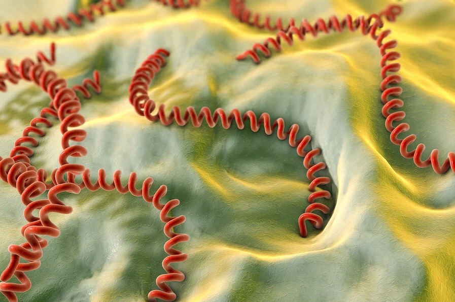
These bacteria, which belong to the genus Leptospira, cause leptospirosis, a disease that affects humans and animals (including dogs, cattle and horses), for which there are currently no effective vaccines. They are close relatives of other pathogens such as Treponema pallidum, the bacterium that causes syphilis — a sexually transmitted disease for which there is also no effective vaccine available. Together, they belong to the group known as spirochaetes: bacteria with a spiral-shaped body, like a corkscrew.
Tiny motors, extraordinary speeds
Many bacteria move thanks to flagella, long, thin appendages made up of thousands of copies of the same protein that rotate like a motor propeller. Some can reach remarkable speeds: scaled up to human size, this would be equivalent to swimming 10 or 15 times faster than an Olympic swimmer.
In Leptospira, things are different: their flagella are located inside the cell, between the outer membrane and the bacterial body, which is why they are called endoflagella. Even so, they rotate, and this rotation is essential for the bacterium to swim efficiently and infect its host.
A system more complex than expected
The study by the Molecular and Structural Microbiology Laboratory at IP Montevideo, published in Nature Communications, describes for the first time, in atomic detail, how the Leptospira endoflagellar filament is built.
Using high-resolution electron microscopy (cryo-electron microscopy), the team found that the filament is not made up of a single protein repeated thousands of times, as is the case in most bacteria, but of many different proteins. The typical flagellar protein is still present, but it forms only the central core, which is completely surrounded by a dozen different proteins arranged very precisely around it, like a glove covering a hand. To solve this genuine puzzle, the team took advantage of other important techniques to measure proteins and determine whether they are close to their neighbours, as well as to modify the bacteria’s genetic information and investigate their molecular functions.
The filament’s architecture determines how the bacterium swims
One of the key findings is that the proteins in the core “choose” their partners in the surrounding sheath. Leptospira can build different types of core because it has four variants of the flagellar protein (flagellins), and this interaction between the core and the sheath determines the curvature of the filament.
The team showed that pathogenic species (those that cause leptospirosis) have less curved filaments than free-living, non-pathogenic species. And this difference is significant: because they are less curved, pathogenic species swim much more effectively through viscous environments, such as those they must navigate inside an infected organism.
To test this, the researchers genetically modified Leptospira interrogans (a pathogenic species) so that the core of its filament was formed using the typical flagellin of a non-pathogenic species. This change caused the sheath to reorganise and the filament to become more curved. As a result, although these modified bacteria grew normally, they completely lost their ability to cause infection: they could no longer swim effectively through viscous media or move through tissues.
Towards new prevention strategies
Beyond revealing a unique mechanism of movement among bacteria, understanding how this system — which is essential for infection — is built opens the door to identifying molecular targets for the development of vaccines against leptospirosis, and perhaps, in the future, against other diseases caused by spirochaetes.
This research was led by Fabiana San Martín (whose doctoral thesis at PEDECIBA focused on these studies), together with Sonia Mondino, Nicole Larrieux, Felipe Trajtenberg, Alejandro Buschiazzo, Azalia Rodríguez and Rosario Durán, from IP Montevideo, in collaboration with scientists from the United States and France.
